
Client: Johns Hopkins Graduate Student - Baltimore, MD
Project: Thesis Research Graphics
Role: Graphic Design, Illustration
Tools: Pencil & Paper, Photoshop, Affinity Designer

The client in this case is a graduate student who required graphics for scientific publications and presentations. The graphics I designed were for several projects within the realm of fusion protein design and construction. Fusion protein production involves the use of genetic engineering techniques to “fuse” two or more gene fragments that are associated with unique functions. The combination of such entities results in a new and synthetic fusion protein that has a hybrid function (the function relating to the fragments it was constructed from). During this project, I was tasked with visually communicating complex scientific concepts with an emphasis on accuracy, clarity, and simplicity.
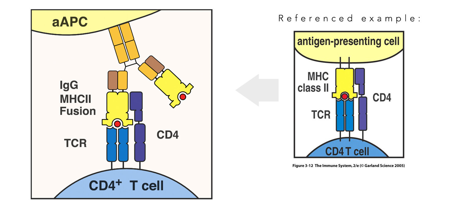
On the left we see an example of an MHC II/antibody fusion protein engaging and activating a T cell. On the right we see the conventional diagram for T cell engagement by MHC II (Garland Science 2005), which I referenced in order to model the fusion protein according to the client’s specifications. Scientific language is a highly developed form of dialect intended to communicate biological elements to other scientists by using agreed upon symbols and shapes that may seem arbitrary to a layman but are highly recognizable to a scientist. For instance, notice the shape and number of domains on the MHC II vs the antibody vs the TCR surface receptor—these graphic elements communicate to a scientist the identity of these molecules even in absence of labeling.
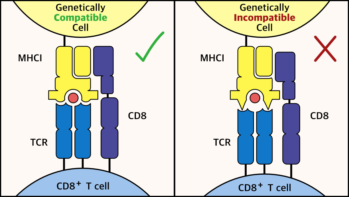
Here we see a diagram explaining the mechanism behind graft rejection, mediated by shape incompatibility between an MHCI and a TCR. On the left we see the lock and key interaction between MHC I and TCR, resulting in recognition of “self” cells; while on the right we see recognition of a cell as foreign due to biophysical incompatibility between these two molecules. To achieve visual separation between the two cell types, I chose colors around the yellow spectrum for molecules belonging to the top cell and colors around the blue spectrum for molecules belonging to the T cell.
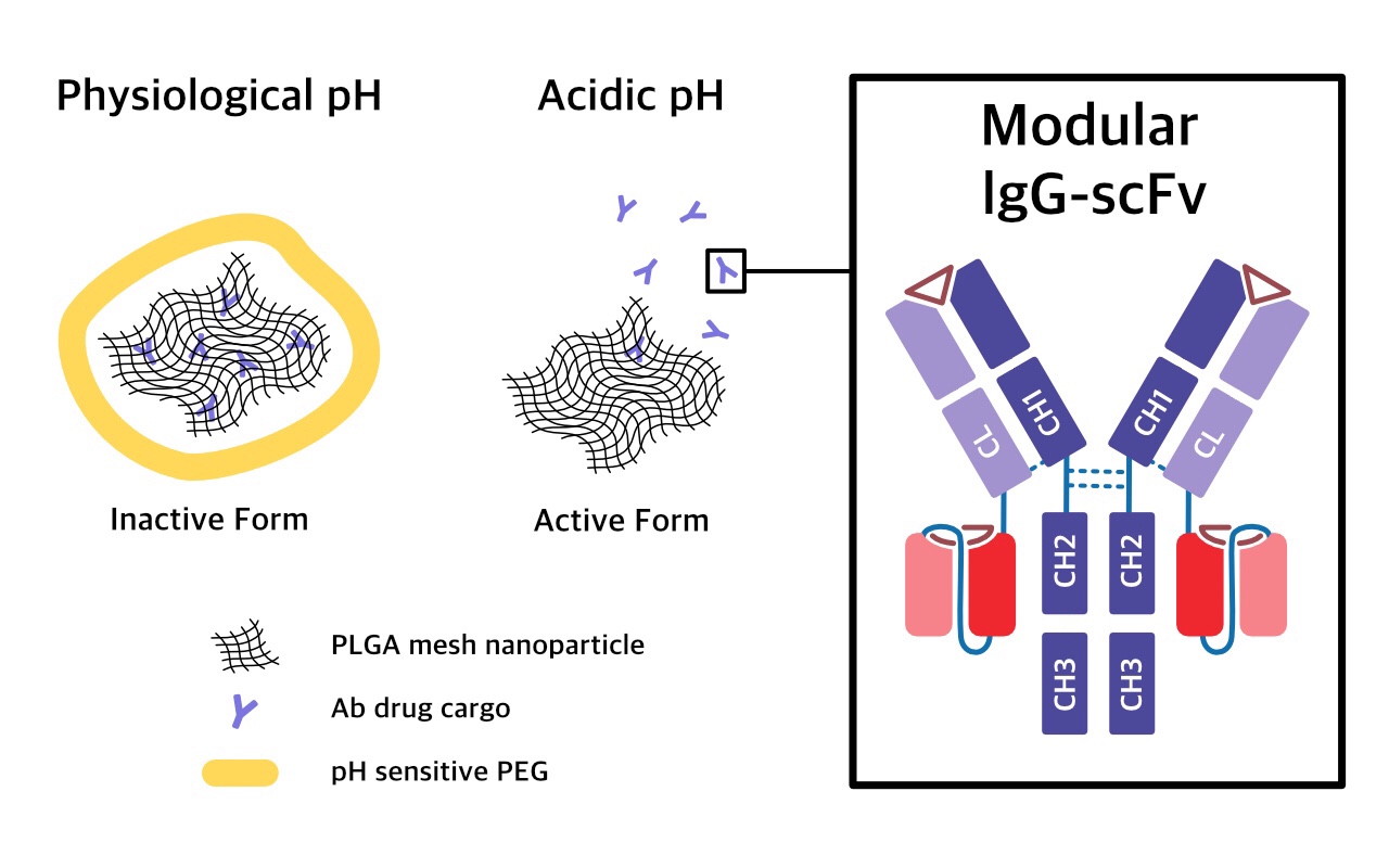
This diagram portrays a tumor targeting drug delivery vehicle carrying a bispecific antibody therapeutic. The drug carrier is coated with PEG which is sensitive to the acidic tumor microenvironment, resulting in the degradation of PEG and release of drug selectively in the tumor. It was important to convey the drug release mechanism clearly without overwhelming the image with unnecessary detail. In addition, the accuracy when representing elements in this image was important to the client. For instance, where a nanoparticle would typically be depicted as a circle—the structure of PLGA nanoparticles is actually mesh. Notice the blue linker between the CL domain and scFv, this linker accurately represents the manner in which the antibody domains were linked.
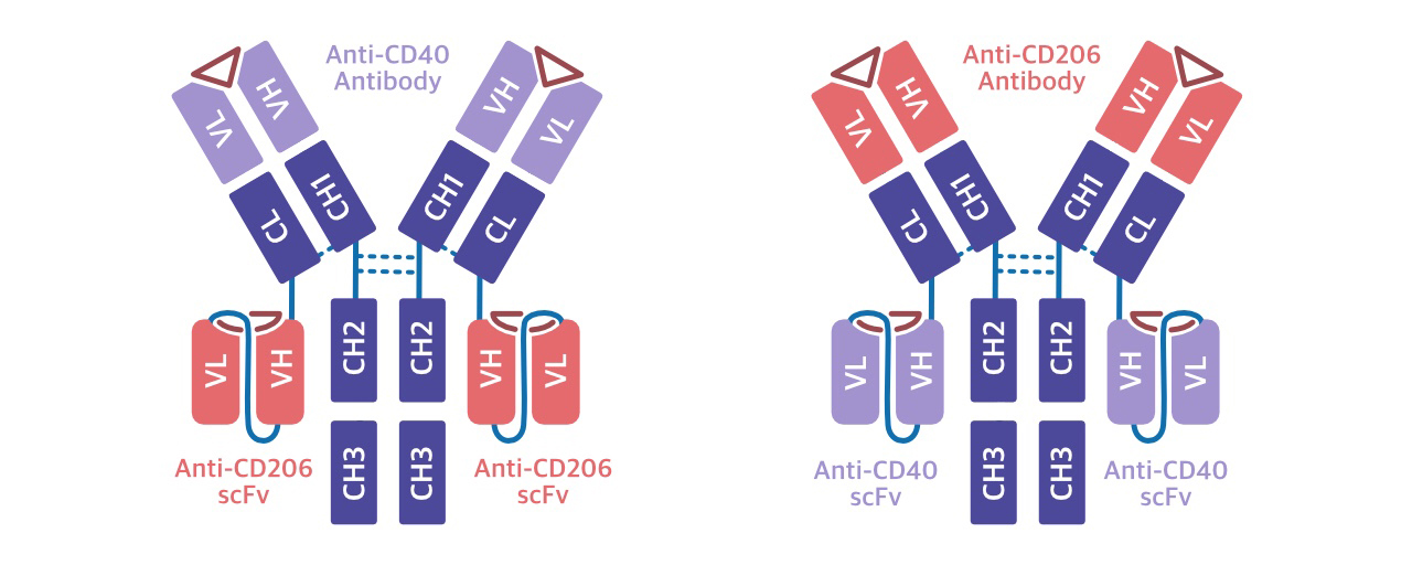
Graphic depicting two different formats for a bispecific antibody.
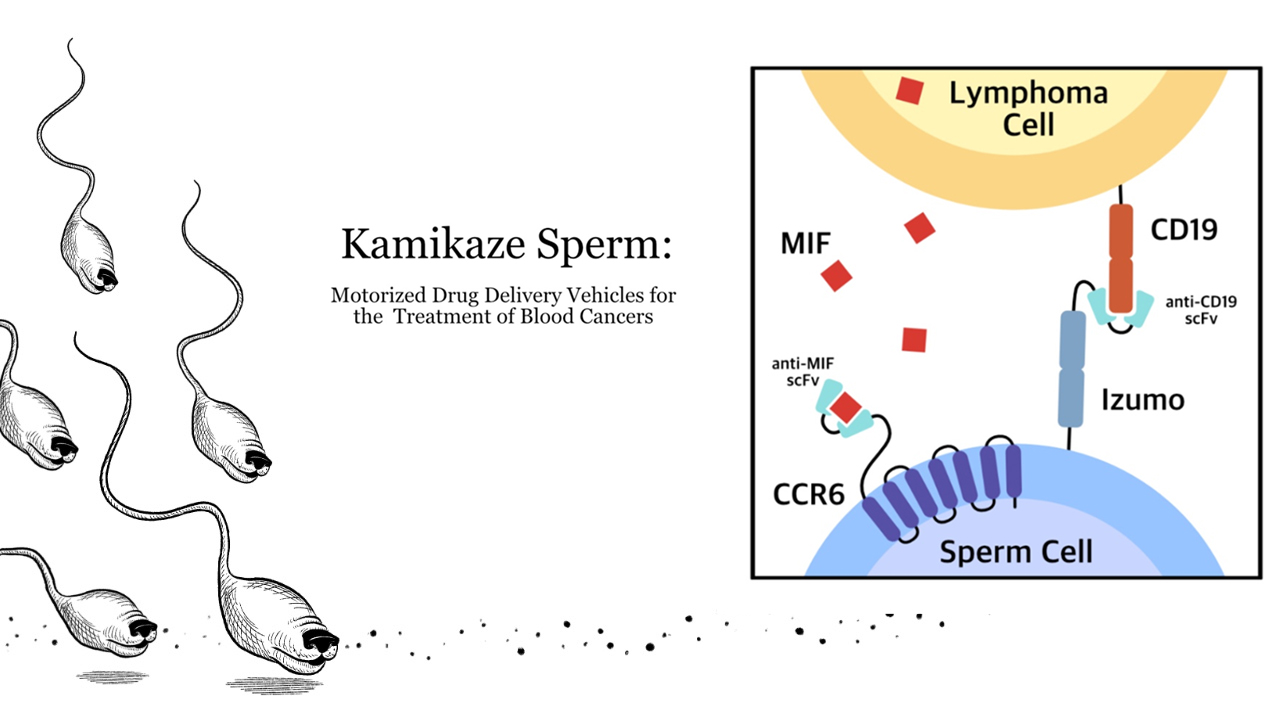
For this project, sperm cells were engineered as drug carrying homing mechanisms that can track and attack cancer cells. I wanted to leverage my skills as an illustrator to depict these kamikaze sperm in a humorous way. My client used the illustration on the left as the title slide for the presentation, the cancer tracking “hound sperms” successfully disarmed the audience. The diagram to the right depicts the two fusion receptors that mediate the interaction between the engineered sperm cell and the cancer cell. Again, the shapes and color choice were very purposeful, conveying both accuracy and visual organization. For instance, CCR6 is a seven-pass transmembrane receptor and as such should be graphically depicted as having seven membrane domains. Due to my client’s creativity and partially due to the success of my graphics, the presentation was voted first place out of dozens of student presentations.
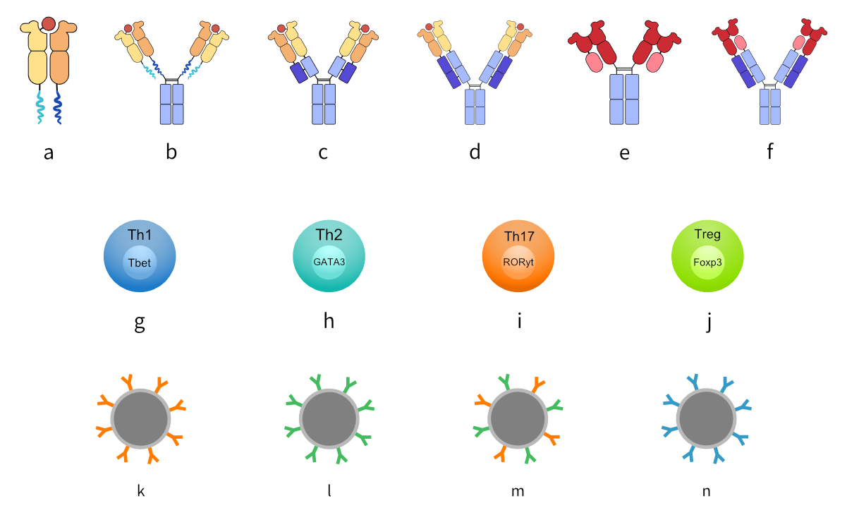
Here we see vector assets for my client’s thesis project, including a selection of fusion protein formats. Since these proteins are synthetic and therefore graphically non-referenceable, the client needed my help to communicate protein design decisions and to showcase the variety of different formats she engineered. (a-f) are MHC fusion proteins, with MHCs in warm colors and dimerization domains in cool colors. (g-j) represent different types of T cells, each phenotype is coupled with its associated transcription factor. (k-n) depict nanoparticles mounted with different antibodies.
